Retinal Vein Occlusion — Branch (BRVO) and Central (CRVO)
Retinal Vein Occlusion — Branch (BRVO) and Central (CRVO)
Retinal vein occlusion is one of the few moments in medicine where the Western structural diagnosis and the classical Chinese diagnosis are, at their core, the same statement. The central retinal vein closes. A branch retinal vein closes. The blood upstream of the closure cannot drain. Pressure builds behind the obstruction. The vessel walls, unable to contain the hydrostatic load, give way. Blood escapes into the retina. Fluid accumulates at the macula. The photoreceptors that depend on that fluid space being clear are buried under the edema. Vision blurs, darkens, or drops.
血瘀阻絡。 Blood stasis obstructs the collaterals.
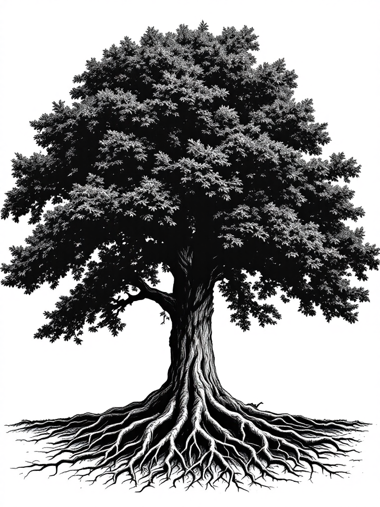
This is the foundational classical statement for RVO. The retinal vessels — particularly the fine venous drainage channels — are collateral vessels in the classical anatomy: the secondary distribution channels that carry Blood from the tissues back to the primary circulatory network. When Blood stasis accumulates in those collaterals, the flow slows, thickens, and eventually stops. The Western mechanism is thrombosis at a specific anatomical chokepoint: the lamina cribrosa for central retinal vein occlusion, where the optic nerve and retinal vein share a confined passage through the scleral canal; the arteriovenous crossing point for branch occlusion, where a sclerotic retinal artery compresses the vein beneath it. The classical mechanism is Blood stasis in the collateral, which is the same event described in a physiological language that also captures why that patient's Blood became stagnant, why those particular collaterals were vulnerable, and what the constitutional terrain was that made the thrombosis possible.
The two other classical statements that complete the foundational picture:
脾統血。 The Spleen holds Blood within the vessels.
When the Spleen fails in its governing function, Blood does two things simultaneously: it escapes the vessels (hemorrhage) and it stagnates within them (thrombosis). This is not contradictory — it is a precise description of what happens in RVO. The flame hemorrhages that characterize both BRVO and CRVO are Blood escaping because the Spleen's holding function is insufficient. The occlusion that caused the hemorrhages is Blood stagnating because the Spleen's transformative and circulatory support has failed. Both events, in the classical framework, have the same upstream driver: Spleen Qi has failed to govern the Blood.
久病入絡。 Chronic disease enters the collaterals.
The third governing statement applies to every RVO patient with a history of hypertension, hyperlipidemia, diabetes, or atherosclerotic vascular disease. Long-standing Blood stasis — years of thickened, poorly circulating Blood driven by metabolic dysregulation — accumulates progressively in the finest collateral channels. The retinal veins are among the smallest and most geometrically constrained vessels in the body. They receive the accumulated effect of years of constitutional Blood stasis before a Western diagnosis of vascular disease has been made. When the accumulated stasis reaches a critical threshold at the lamina cribrosa or the arteriovenous crossing, the vein occludes. The thrombosis is the terminal event in a progression that the classical framework had been tracking for decades.
These three statements — Blood stasis in the collaterals, Spleen failing to hold Blood, chronic stasis entering the collaterals — form the classical architecture for RVO. They do not contradict the Western pathophysiology. They explain the constitutional substrate that the Western pathophysiology requires in order to occur.
Western retinal vascular medicine has mapped the structural sequence of RVO in precise detail. The anatomy creates the vulnerability: the central retinal vein and the central retinal artery share the same tight passage through the lamina cribrosa, a sieve-like plate of connective tissue at the optic nerve head. In CRVO, a thrombus forms at this passage — typically at a point of arteriovenous contact where the artery wall, stiffened by atherosclerosis, compresses the venous lumen and disturbs laminar flow enough to activate the coagulation cascade. In BRVO, the same mechanism operates at one of the downstream arteriovenous crossing points in the peripheral retina, where a branch retinal artery crosses directly over a branch retinal vein and compresses it.
Once the vein occludes, three parallel processes begin:
Venous drainage obstruction. The entire retinal territory drained by the occluded vein — half the retina in a major BRVO, the entire retina in CRVO — loses its outflow pathway. Hydrostatic pressure rises throughout the venous network. The retinal capillary bed, unable to drain, becomes congested. The characteristic flame hemorrhages of RVO are the result: blood forced through the congested capillary walls into the nerve fiber layer, following the direction of the axons in a pattern so distinctive it is immediately recognizable on funduscopy.
Macular edema. The macula — the central retinal zone responsible for high-acuity vision — is particularly sensitive to venous congestion. Fluid accumulates in the outer nuclear layer and the spaces between the photoreceptors, forming cystoid macular edema. The photoreceptors are displaced and their function is compromised. Central vision blurs. In CRVO, macular edema is the primary mechanism of vision loss and is frequently the most refractory aspect of the condition to manage. Anti-VEGF therapy — intravitreal injection of bevacizumab, ranibizumab, or aflibercept — targets the vascular endothelial growth factor that is driving the edema, but the injections must be continued monthly in many patients for months to years because the underlying occlusion remains unresolved.
Retinal ischemia and neovascularization. When the capillary non-perfusion is extensive — in ischemic CRVO particularly — the hypoxic retina upregulates VEGF production as part of the angiogenic response to oxygen deprivation. New vessels grow: at the optic disc, at the retinal surface, and eventually at the anterior segment iris and angle (rubeosis iridis). These new vessels are fragile, poorly structured, and bleed easily. Vitreous hemorrhage from neovascular rupture can cause sudden, severe vision loss on top of the existing macular edema. Neovascular glaucoma — new vessels growing across the trabecular meshwork and blocking aqueous outflow — is the most serious structural complication of ischemic CRVO.
The risk factors that the Western literature identifies for RVO are the same constitutional variables the classical framework identifies as the drivers of long-standing Blood stasis: hypertension (Liver Yang rising, internal pressure dysregulation); hyperlipidemia (Phlegm-Blood stasis — thick, sluggish Blood unable to circulate freely through fine channels); diabetes (Phlegm-Dampness and Blood stasis combined); hyperviscosity states including Waldenström's macroglobulinemia and multiple myeloma (extreme Blood thickening — the classical picture of extreme Phlegm-Blood stasis); thrombophilia including Factor V Leiden and antiphospholipid syndrome (constitutional Blood that clots too easily — the classical picture of chronic constitutional Blood stasis with a heat or cold driver maintaining the tendency); and glaucoma, which compresses the optic nerve head including the lamina cribrosa through which the central retinal vein passes.
久病入絡。 Chronic disease enters the collaterals.
Every item on that risk factor list is a classical description of long-standing Blood stasis progressively entering the fine collaterals. The RVO is not the beginning of the disease. It is the thrombotic terminus of a constitutional process that has been accumulating for years, visible in the pulse, the tongue, and the systemic pattern long before a vein in the retina closes.
Each classical statement names a physiological relationship. Each relationship, when it fails, produces a predictable Western finding. The bridge between them is not metaphorical — it is mechanistic, and reading the table in both directions shows why the same Western diagnosis of RVO arises from constitutionally distinct upstream drivers that require different formulas.
This section is provided as clinical reference. Classical Chinese medical statements and their pattern mechanisms are described below, not disease claims. All formula recommendations represent classical pattern-based support, not treatment of diagnosed disease conditions.
| Western Finding | Classical Statement | Pattern | Formula Direction |
|---|---|---|---|
| Retinal vein thrombosis | 血瘀阻絡 Blood stasis obstructs collaterals |
Blood Stasis (primary) | Xue Fu Zhu Yu Tang 血府逐瘀汤 |
| Macular edema | 脾失運化 Spleen fails to transform and move fluid |
Spleen Qi Deficiency + Fluid Stagnation | Bu Zhong Yi Qi Tang + Ze Xie 泽泻 (drain fluid accumulation) |
| Flame hemorrhages | 脾統血 Spleen fails to hold blood in the vessels |
Spleen Qi Deficiency + Blood Stasis | San Qi 三七 — moves existing stasis AND arrests hemorrhage simultaneously |
| Hypertension root (CRVO driver) | 肝氣鬱結 Liver Qi stagnation → Yang rising |
Liver Qi Stagnation converting to Heat | Dan Zhi Xiao Yao San 丹栀逍遥散 |
| Hyperlipidemia / hyperviscosity | 痰瘀互結 Phlegm and Blood stasis combine |
Phlegm-Blood Stasis | Wen Dan Tang + Dang Gui 温胆汤加当归 |
| Ischemia → neovascularization (VEGF upregulation) | 久病入絡 Chronic disease enters the collaterals |
Long-standing stasis → ischemic collateral invasion | XFZYD + San Qi — move stasis, arrest hemorrhage, protect remaining perfused tissue |
The table makes the clinical point visible: RVO is not a single-pattern disease. The thrombosis is the shared event. The constitutional terrain that made it possible — the specific upstream pattern of Blood stasis, Qi deficiency, Liver constraint, Phlegm accumulation, or Yang deficiency — varies from patient to patient. Two people walk out of the retinal specialist's office with the same CRVO diagnosis. Their formulas will not be the same. The lock that produced the occlusion is different in each case, and the key must be cut for the specific lock.
Chinese herbal formulas are chemical interventions. The four actions on Blood — move, cool, generate, warm — describe measurable pharmacological effects on coagulation dynamics, inflammatory signaling, vascular tone, erythrocyte deformability, platelet aggregation, and tissue nutrition. In RVO, the proportioning of these four actions is dictated by the constitutional pattern. MOVE is primary in nearly every RVO case because the fundamental lesion is Blood stasis. The other three actions are added, proportioned, and sequenced according to what pattern produced the stasis.

MOVE the Blood (primary action — overwhelmingly a Blood-stasis condition). Every RVO formula contains a Blood-moving core. The question is which movers, in what proportion, and what else is added around them.
COOL and MOVE (Liver-heat and Blood-stasis overlap). In the hypertension-driven RVO patient — stressed, constitutionally warm, wiry pulse — the Blood stasis is occurring in a vascular environment that is already hot and pressurized. Heat thickens the Blood and drives it upward toward the head and eyes. Moving Blood into a heat environment without cooling first is less effective; the cooling action creates the vascular environment in which the movers can work.
GENERATE Blood (recovery and ischemia phase). The hemorrhages of RVO represent Blood lost from the vessels into the retinal tissue. Ischemia means the photoreceptors and retinal ganglion cells are not receiving adequate perfusion. The Blood that was lost must be rebuilt; the tissue that is starved must be nourished. GENERATE is the recovery action — it follows MOVE in the sequence, or runs in parallel at a lower dose when Blood deficiency is evident at presentation.
WARM (cold-stasis pattern, elderly or Yang-deficient constitution). In the elderly, cold-constitution RVO patient — low energy, pale tongue, slow pulse, cold extremities — Blood stasis is occurring in a circulatory environment that lacks the Yang warmth needed to drive Blood through the fine retinal channels. Heat moves Blood; cold stagnates it. Cold-stasis RVO requires warming alongside moving, or the Blood-movers work against a constitutional current that is slowing everything down.
Representative formulas that deploy these four actions in different configurations for RVO:
Two patients present to the same retinal specialist on the same afternoon. Both have CRVO. Both have macular edema on OCT. Both are scheduled for monthly anti-VEGF injections. Their Western treatment protocol is identical. Their classical formulas will not be.

The first patient is fifty-five years old. He is a driven, high-functioning professional — the kind of person who sleeps four hours, drinks three espressos before noon, and describes himself as "always on." His face is slightly flushed. His blood pressure has been borderline elevated for years; his cardiologist has been watching it without prescribing. He gets tight headaches at the temples. His tongue is red with a thin yellow coat. His pulse is wiry and slightly rapid. He came to RVO from the classic hypertension pathway: Liver Qi stagnation accumulated over years of constraint and stress; stagnation converted to heat; Liver Yang rose; internal vascular pressure elevated; the central retinal vein at the lamina cribrosa — already compressed by disc anatomy — gave way under the hydrostatic load. His combination lock opens: first cool the Liver heat, descend the rising Yang, remove the vascular pressure environment that produced the thrombosis — Dan Zhi Xiao Yao San with Chi Shao and Mu Dan Pi. Then move the stasis — XFZYD layered in as the heat pattern begins to resolve. San Qi throughout, because his flame hemorrhages are still absorbing. Not Bu Yang Huan Wu Tang. He does not have Qi deficiency driving cold stasis; he has heat driving Blood stasis. Warming herbs would add fuel to the wrong fire.
The second patient is sixty-eight years old. She is a retired teacher — quiet energy, speaks slowly, gets cold easily. Her blood pressure is normal. She has no history of stress-related illness. She has had low energy for years, her digestion is unreliable, and she has been slowly gaining weight despite eating modestly. Her tongue is pale and slightly swollen at the edges. Her pulse is deep, slow, and slightly slippery. Her RVO came not from Yang excess driving pressure upward but from Yang deficiency failing to warm and circulate Blood through the smallest channels — a cold-stasis RVO in a Qi- and Yang-deficient constitution. Her combination lock opens differently: warm and move — Bu Yang Huan Wu Tang with Gui Zhi, Dang Gui generating Blood alongside the moving herbs, San Qi for the hemorrhage component. Her macular edema has a fluid-stagnation component from Spleen failing to transform fluid; Ze Xie is added to drain the fluid accumulation while the primary formula addresses the cold-stasis root. No strong heat-clearing herbs. She does not have a heat pattern. Cooling her further would deepen the constitutional stasis that drove the occlusion in the first place.
The combination lock variables in RVO:
一人一方。 One person, one formula.
The formula follows the pattern. The pattern follows the person. The intake exists to establish which lock is in front of us before the key is cut.
RVO patients are rarely thinking about their digestion. They are thinking about the vision that dropped this week. The retina is where the attention goes, appropriately. But the classical framework has a governing rule about formula delivery that the retinal focus does not override:
脾胃者,後天之本。 The Spleen and Stomach are the root of post-natal life.
The Spleen and Stomach generate the Qi and Blood from which every other tissue — including the retina, the macular photoreceptors, the retinal ganglion cells, and the peripapillary capillary network — is nourished and sustained. If the Spleen's transformative function is impaired, the Blood generated from food is insufficient in quality and volume; the herbs in the formula move through a compromised digestive infrastructure; and the ischemic retinal tissue that the formula is designed to nourish is not receiving the delivery that should be reaching it.
This matters specifically in RVO because the macular edema that is destroying central vision is, in the classical framework, partly a Spleen failure: 脾失運化 — the Spleen has failed to transform and move fluid. The fluid accumulates at the macula not only because the venous drainage is obstructed but because the Spleen is no longer doing its metabolic work of circulating and resolving fluid accumulation throughout the body. Anti-VEGF injections reduce VEGF-driven vascular permeability; they do not address the constitutional fluid metabolism failure that is contributing to the edema from the systemic side.
The functional medicine parallel maps precisely: before loading the liver-intensive chemistry of a full Blood-moving formula, you assess whether the gut's absorptive infrastructure is intact. San Qi, Dan Shen, Dang Gui, and Chi Shao need to be broken down, absorbed through the intestinal epithelium, packaged in appropriate lipid carriers, transported to the hepatic portal system, metabolized to their active constituents, and delivered to the retinal microcirculation. Each step in that chain requires a functional digestive system. If the patient has chronic bloating, irregular bowel function, poor appetite, or food sensitivity — classical Spleen Qi deficiency signs — those herbs are moving through a compromised delivery network, and the formula is less effective than its chemistry should make it.
Where Spleen Qi support is indicated — and in RVO patients with macular edema and constitutional digestive weakness, it usually is — Dang Shen (Dǎng Shēn, 党参), Bai Zhu (Bái Zhú, 白术), and Fu Ling (Fú Líng, 茯苓) are incorporated into the formula to build the transformative capacity that delivers everything else. Ze Xie (Zé Xiè, 泽泻) drains the fluid accumulation that compromised Spleen function is permitting. The Blood-moving core of the formula works through a system that can actually process and deliver it.
Here is what the monthly anti-VEGF appointment looks like from inside it: you sit in a waiting room. A retinal specialist dilates your pupil, confirms macular edema on OCT, injects bevacizumab or aflibercept into your vitreous — a needle into the white of the eye, which is as uncomfortable as it sounds — and books you for the same thing in four weeks. Each appointment costs between $1,500 and $2,500. Many patients continue for twelve to twenty-four months. Some indefinitely. The injections work; they reduce VEGF-driven vascular permeability, the edema resolves on OCT, and central vision recovers partially or substantially in many patients. Continue them. This is not an argument against them.
This is an argument about what the injections do not address.
Anti-VEGF therapy manages the consequence of the occlusion: the macular edema driven by VEGF upregulation in the ischemic retinal tissue. It does not address the Liver Qi stagnation that elevated your internal vascular pressure for fifteen years before the vein closed. It does not address the Blood stasis constitution that thickened your Blood and slowed your retinal circulation until the thrombosis was inevitable. It does not address the Phlegm-Blood stasis of hyperlipidemia accumulating in your retinal vessels across the decade before the occlusion. It does not address the Spleen Qi deficiency that is allowing fluid to accumulate at your macula from the systemic side, compounding the VEGF-driven permeability the injection is suppressing. And it does not address the risk of the next occlusion — in the same eye, in the fellow eye, or in another vascular territory — because the constitutional terrain that produced the first one is still present and operating.
The statistics on fellow-eye involvement in RVO are not reassuring. Risk of contralateral RVO in the years following an index event is meaningful, particularly in patients with systemic vascular risk factors. The fellow eye shares the same constitutional Blood-stasis environment that occluded the first vein. Nothing in the anti-VEGF protocol addresses that shared constitutional terrain.
The classical herbal framework addresses the lock, not just the hole in the vision. It reads the upstream constitutional pattern — the heat-cold axis, the Qi-deficiency driver, the Phlegm accumulation, the Liver Qi constraint — and formulates directly to that pattern. You are not choosing between the injection and the formula. You are running both: ophthalmology manages the structural consequence in the eye it can see; the formula addresses the constitutional terrain that produced the occlusion and that remains capable of producing the next one.
The injection keeps the edema suppressed while the formula changes the soil. Both are necessary. Neither replaces the other. The question is whether you address only what happened, or also what made it possible.
The online intake gathers the full clinical picture: your Western diagnosis (BRVO or CRVO, which eye, how long ago the occlusion occurred), your most recent OCT findings including central macular thickness, your current anti-VEGF injection schedule if you are in one, your systemic risk factors (hypertension, hyperlipidemia, diabetes, any thrombophilia diagnosis), and your current medications. All of this maps the Western context that the formula design must work within.
The constitutional intake goes deeper: the heat-cold axis of your constitution; your energy levels and how they have changed since the occlusion; your digestive function; your sleep and stress patterns and their relationship to your vascular history; whether you have any history of other vascular events; what your most recent bloodwork showed for lipids, glucose, homocysteine, and inflammatory markers; and the full subjective picture of your vision — what has recovered, what has not, how your central vision compares to your pre-occlusion baseline.
This information maps the classical pattern: which driver produced the stasis, whether heat or cold or deficiency is the constitutional context, what Spleen support is needed for the formula to be delivered effectively, and what stage of the disease progression you are in — acute, resolving, chronic-edema, or recovery phase. The formula is designed from that map.
Michael reads every intake personally, identifies the primary and secondary patterns, determines the formula architecture and the sequence in which it will shift as the pattern responds, and sends a written explanation of the formula's rationale with your first shipment so you understand what you are taking and why.
If you are currently receiving anti-VEGF injections, continue them without interruption. The formula does not replace intravitreal therapy for active macular edema. It addresses the constitutional terrain that the injections do not reach. Some patients find, over the course of herbal support, that injection intervals extend as the constitutional pattern improves; this should be assessed and confirmed by your retinal specialist on OCT, not assumed. Communicate with your retinal specialist that you are using herbal support so that the full clinical picture can be considered at each monitoring appointment. The formula and the injection protocol are complementary. They address different layers of the same condition.
The Chambers are a free patient education library — the methodology behind every Rootworth formula. Reading them before or alongside your intake helps you understand what the classical assessment is seeing, why individualized formulas outperform generic protocols, and how each layer of treatment connects to the next.
A note on these statements.
Rootworth herbal preparations are dietary supplements. These statements have not been evaluated by the Food and Drug Administration. These products are not intended to diagnose, treat, cure, or prevent any disease. Classical Chinese medicine pattern assessment — the identification of constitutional patterns such as Blood stasis, Spleen Qi deficiency, Liver Qi stagnation, Phlegm-Blood stasis, or Kidney Yang deficiency — is distinct from the diagnosis and treatment of disease as defined under United States federal law. Individual results vary. All formula recommendations on this page represent classical pattern-based herbal support and are not claims to treat, manage, or cure retinal vein occlusion, macular edema, retinal ischemia, or any associated ocular or systemic condition. Always continue your retinal specialist's monitoring and any prescribed treatments alongside any herbal support program. Do not interrupt intravitreal injection schedules or alter any ophthalmologist-prescribed care without the direct guidance of your retinal specialist.